Lisfranc Injury Mri : Lisfranc Injury Radiology Reference Article Radiopaedia Org - Mri online is a premium online continuing education resource for practicing radiologists to expand their radiology expertise across all modalities, read a wide variety of cases, and become a more accurate, confident, and efficient reader.
Lisfranc Injury Mri : Lisfranc Injury Radiology Reference Article Radiopaedia Org - Mri online is a premium online continuing education resource for practicing radiologists to expand their radiology expertise across all modalities, read a wide variety of cases, and become a more accurate, confident, and efficient reader.. 4,15 mr imaging has been shown to demonstrate the lisfranc ligament complex reliably in the normal foot. It can range from mild to severe. Learn how to identify the lisfranc ligament complex and the bifurcate ligament on mri. Of the lisfranc complex when adding a specific mri study for this type of injury to the process of diagnosing subtle lisfranc injuries. Mri findings as many as 20 percent of lisfranc joint injuries are missed on initial anteroposterior and oblique radiographs.
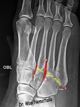
Even though recent advances in mri techniques have improved the specificity for the diagnosis of injuries to the different ligaments of the lisfranc complex, the distinction between the lisfranc ligament and the nearby plantar ligaments has not always been clearly made in the past. Lisfranc joint injuries are relatively uncommon, and their imaging findings can be subtle. The tarsometatarsal, or lisfranc, joint complex provides stability to the midfoot and forefoot through intricate osseous relationships between the distal tarsal bones and metatarsal bases and their connections with stabilizing ligamentous support structures. Magnetic resonance imaging is accurate for detecting traumatic injury of the lisfranc ligament and for predicting lisfranc joint complex instability when the plantar lisfranc ligament bundle is used as a predictor. 17 three distinct structures can be identified:
Lisfranc Injury Radiology Reference Article Radiopaedia Org from prod-images-static.radiopaedia.org Summary a lisfranc injury is a tarsometatarsal fracture dislocation characterized by traumatic disruption between the articulation of the medial cuneiform and base of the second metatarsal. Most mri studies assessed lisfranc ligament integrity. Learn how to identify the lisfranc ligament complex and the bifurcate ligament on mri. Injuries to the tarsometatarsal joint can be caused by low or high impact. Mri findings as many as 20 percent of lisfranc joint injuries are missed on initial anteroposterior and oblique radiographs. Typically must injure lisfranc ligament and plantar ligament from first cuneiform and 2nd and 3rd mt bases proximal or medial column variant seen more often in high level football players. Lisfranc joint injuries are relatively uncommon, and their imaging findings can be subtle. You will learn that the dorsal lisfranc ligament is the weakest and th.
Even though recent advances in mri techniques have improved the specificity for the diagnosis of injuries to the different ligaments of the lisfranc complex, the distinction between the lisfranc ligament and the nearby plantar ligaments has not always been clearly made in the past.
Lisfranc (midfoot) injury lisfranc (midfoot) injuries result if bones in the midfoot are broken or ligaments that support the midfoot are torn. 17 three distinct structures can be identified: Your foot will likely also be unable to bear weight. Of the lisfranc complex when adding a specific mri study for this type of injury to the process of diagnosing subtle lisfranc injuries. Summary a lisfranc injury is a tarsometatarsal fracture dislocation characterized by traumatic disruption between the articulation of the medial cuneiform and base of the second metatarsal. For the study, a 1.5 tesla mri has been used to perform the exams, placing the patient in a comfortable position with a suitable coil, looking for an immobilized forefoot but without Therefore, no imaging reference can be used for related diagnosis and repair operations. Hence, there is no available auxiliary examination for diagnosing related injuries. Your lisfranc joint injury might cause bruising, deformity, swelling, or pain in the middle of your foot. Typically must injure lisfranc ligament and plantar ligament from first cuneiform and 2nd and 3rd mt bases proximal or medial column variant seen more often in high level football players. Lisfranc joint injuries are relatively uncommon, and their imaging findings can be subtle. Diagnosis is confirmed by radiographs which may show widening of the interval between the 1st and 2nd ray. Force extends through intercuneiform joint of medial and middle cuneiforms and
6,13,17 on mri, acute ligamentous injury appears as alterations in normal ligament signal and. There were associated comminuted fractures of head of 2 nd metatarsal, phalanges of 2 nd digit with dislocation of 2 nd metatarsophalangeal joint. 4,15 mr imaging has been shown to demonstrate the lisfranc ligament complex reliably in the normal foot. The tarsometatarsal, or lisfranc, joint complex provides stability to the midfoot and forefoot through intricate osseous relationships between the distal tarsal bones and metatarsal bases and their connections with stabilizing ligamentous support structures. Mri findings as many as 20 percent of lisfranc joint injuries are missed on initial anteroposterior and oblique radiographs.
Subtle Lisfranc Injury In Mri Specific Protocol For A Detailed Report Semantic Scholar from d3i71xaburhd42.cloudfront.net 4,15 mr imaging has been shown to demonstrate the lisfranc ligament complex reliably in the normal foot. Diagnosis is confirmed by radiographs which may show widening of the interval between the 1st and 2nd ray. However, in clinically suspected cases of traumatic lisfranc ligament injury, true positive rate for sprain is low. He loves discussing and writing about sports & exercise injuries, and has been featured in major media publications over 1,200 times throughout his career. A lisfranc injury occurs when one or more of the metatarsal bones are displaced from the tarsus, which is a cluster of bones at the top of the foot, just below the ankle joint. Lisfranc injury may range from isolated ligamentous sprain to complete disruption of the tarsometatarsal articulation. It can range from mild to severe. Of the lisfranc complex when adding a specific mri study for this type of injury to the process of diagnosing subtle lisfranc injuries.
Injuries to the tarsometatarsal joint can be caused by low or high impact.
However, in clinically suspected cases of traumatic lisfranc ligament injury, true positive rate for sprain is low. The severity of the injury can vary from simple to complex, involving many joints and bones in the midfoot. 6,13,17 on mri, acute ligamentous injury appears as alterations in normal ligament signal and. There were associated comminuted fractures of head of 2 nd metatarsal, phalanges of 2 nd digit with dislocation of 2 nd metatarsophalangeal joint. Force extends through intercuneiform joint of medial and middle cuneiforms and Hence, there is no available auxiliary examination for diagnosing related injuries. 4,15 mr imaging has been shown to demonstrate the lisfranc ligament complex reliably in the normal foot. For the study, a 1.5 tesla mri has been used to perform the exams, placing the patient in a comfortable position with a suitable coil, looking for an immobilized forefoot but without It can range from mild to severe. Even though recent advances in mri techniques have improved the specificity for the diagnosis of injuries to the different ligaments of the lisfranc complex, the distinction between the lisfranc ligament and the nearby plantar ligaments has not always been clearly made in the past. Most mri studies assessed lisfranc ligament integrity. The tarsometatarsal, or lisfranc, joint complex provides stability to the midfoot and forefoot through intricate osseous relationships between the distal tarsal bones and metatarsal bases and their connections with stabilizing ligamentous support structures. Therefore, no imaging reference can be used for related diagnosis and repair operations.
Mri online is a premium online continuing education resource for practicing radiologists to expand their radiology expertise across all modalities, read a wide variety of cases, and become a more accurate, confident, and efficient reader. 17 three distinct structures can be identified: Your foot will likely also be unable to bear weight. Most mri studies assessed lisfranc ligament integrity. Ct clarifies tarsometatarsal (tmt) joint alignment and occult fractures obscured on radiographs.
Subtle Lisfranc Injury In Mri Specific Protocol For A Detailed Report Semantic Scholar from d3i71xaburhd42.cloudfront.net Diagnosis is confirmed by radiographs which may show widening of the interval between the 1st and 2nd ray. Mri findings as many as 20 percent of lisfranc joint injuries are missed on initial anteroposterior and oblique radiographs. There were associated comminuted fractures of head of 2 nd metatarsal, phalanges of 2 nd digit with dislocation of 2 nd metatarsophalangeal joint. Lisfranc joint injuries are relatively uncommon, and their imaging findings can be subtle. A lisfranc injury occurs when one or more of the metatarsal bones are displaced from the tarsus, which is a cluster of bones at the top of the foot, just below the ankle joint. A lisfranc injury, also known as lisfranc fracture, is an injury of the foot in which one or more of the metatarsal bones are displaced from the tarsus. Learn more about his background, media appearances, and practice. You will learn that the dorsal lisfranc ligament is the weakest and th.
This study aims to observe and describe the morphology and structure of lisfranc ligaments using magnetic resonance imaging (mri), in order to provide imaging reference for the diagnosis and repair of lisfranc joint injuries.
Injuries to the tarsometatarsal joint can be caused by low or high impact. The tarsometatarsal, or lisfranc, joint complex provides stability to the midfoot and forefoot through intricate osseous relationships between the distal tarsal bones and metatarsal bases and their connections with stabilizing ligamentous support structures. 4,15 mr imaging has been shown to demonstrate the lisfranc ligament complex reliably in the normal foot. Your foot will likely also be unable to bear weight. At present, few studies on the imaging of lisfranc ligaments have been reported, and related imaging data are rare. Diagnosis is confirmed by radiographs which may show widening of the interval between the 1st and 2nd ray. Even though recent advances in mri techniques have improved the specificity for the diagnosis of injuries to the different ligaments of the lisfranc complex, the distinction between the lisfranc ligament and the nearby plantar ligaments has not always been clearly made in the past. A lisfranc injury occurs when one or more of the metatarsal bones are displaced from the tarsus, which is a cluster of bones at the top of the foot, just below the ankle joint. Lisfranc injury may range from isolated ligamentous sprain to complete disruption of the tarsometatarsal articulation. You will learn that the dorsal lisfranc ligament is the weakest and th. David geier is an orthopedic surgeon and sports medicine specialist in charleston, south carolina. Learn how to identify the lisfranc ligament complex and the bifurcate ligament on mri. Lisfranc (midfoot) injury lisfranc (midfoot) injuries result if bones in the midfoot are broken or ligaments that support the midfoot are torn.
Mri findings as many as 20 percent of lisfranc joint injuries are missed on initial anteroposterior and oblique radiographs lisfranc injury. Mri is reasonably accurate at detecting traumatic injury to the lisfranc ligament.


Post a Comment