Brain Anatomy Map / Brain Anatomy Vector Illustration Anatomical Blank Head Organ Structure Stock Vector Image By C Vectormine 299764238 : Brain, bones of cranium, sinuses of the face.
Brain Anatomy Map / Brain Anatomy Vector Illustration Anatomical Blank Head Organ Structure Stock Vector Image By C Vectormine 299764238 : Brain, bones of cranium, sinuses of the face.. The outer portion contains neurons, and the inner area communicates with the cerebral cortex. The following table shows the brain regions from the cortex to the brain stem with their functions and associated disorders in english, german and latin. Can you name these brain structures? Mapping 'imbalance' in brain anatomy across the lifespan. Brain, bones of cranium, sinuses of the face.
Mapping 'imbalance' in brain anatomy across the lifespan. Can you name these brain structures? Brain, bones of cranium, sinuses of the face. There are three major divisions of the brain. The brain pro is a must have app for anyone studying anatomy and is aimed at students with a basic to advanced level of anatomical medical knowledge as well as medical professionals.
Neuroanatomy For The Rest Of Us The Basics Of Brain Anatomy And By Zubair Talib Medium from miro.medium.com The brain stem is located in front of the cerebellum and connects to the spinal cord. Foramina, nasal cavity, paranasal sinuses. See more ideas about brain anatomy, homunculus, brain. Dural venous sinuses, veins, arteries. Let's use a common method and divide the brain into three main regions based on embryonic development: There are three major divisions of the brain. Like the cerebral cortex, it has two hemispheres. The brain pro is a must have app for anyone studying anatomy and is aimed at students with a basic to advanced level of anatomical medical knowledge as well as medical professionals.
The user can manipulate the brain on screen, the app also allows sectioned views and the ability to add custom labels.
Reviewed by john morrison, patrick hof, and edward lein. Anatomy of the head on a cranial ct scan : The allen brain explorer (beta) is an application that allows users to browse multimodal datasets in an annotated 3d spatial framework. The midbrain, pons and medulla oblongata. The minecraft map, brain anatomy, was posted by _master. The forebrain, midbrain and hindbrain. Can you name these brain structures? The fourth edition (following editions in 1992, 1998, 2004) of brain maps: Dural venous sinuses, veins, arteries. The 2d coronal reference atlas is created from a 3d volumetric. Maps & transparency the brain model is. The brain and spinal cord are the two main structures of the central nervous system. This amazing organ acts as a control center by receiving, interpreting, and directing sensory information throughout the body.
The mapping effort subdivided each half of the brain into 180 separate partitions, with 83 of those already well established in the field, and 97 new areas ripe for exploration. The top image shows the four main sections of the cerebral cortex: Reviewed by john morrison, patrick hof, and edward lein. This amazing organ acts as a control center by receiving, interpreting, and directing sensory information throughout the body. The midbrain helps control eye movement and processes visual and.
Becoming Mindful Of The Brain And Its Functions Igea Brain Spine Orthopedics from mltmpgeox6sf.i.optimole.com Let's use a common method and divide the brain into three main regions based on embryonic development: Structure descriptions were written by levi gadye and alexis wnuk and jane roskams. Brain, bones of cranium, sinuses of the face. £14.40 inc vat £12.00 exc vat. The following table shows the brain regions from the cortex to the brain stem with their functions and associated disorders in english, german and latin. The anatomy of the brain is complex due its intricate structure and function. Utilize the model of the human brain to locate the following structures / landmarks for the There are three major divisions of the brain.
The cerebrum, the largest part of the human brain, is associated with higher order functioning, including the control of voluntary behavior.
Anatomy mapper has been online since 2009, and has lead to numerous publications and international collaborations. The fourth edition (following editions in 1992, 1998, 2004) of brain maps: A detailed knowledge of the architectural anatomy of the white matter tracts is paramount, for strategically planning for surgical management of parenchymal brain lesions, such as gliomas. The user can manipulate the brain on screen, the app also allows sectioned views and the ability to add custom labels. This amazing organ acts as a control center by receiving, interpreting, and directing sensory information throughout the body. The 2d coronal reference atlas is created from a 3d volumetric. Finally i ve finished the brain. Foramina, nasal cavity, paranasal sinuses. The mapping effort subdivided each half of the brain into 180 separate partitions, with 83 of those already well established in the field, and 97 new areas ripe for exploration. The researchers discovered that our brain's cortex, or outer mantle, is composed of 180 distinct areas per hemisphere. The brain and spinal cord are the two main structures of the central nervous system. This interactive brain model is powered by the wellcome trust and developed by matt wimsatt and jack simpson; The midbrain helps control eye movement and processes visual and.
Let's use a common method and divide the brain into three main regions based on embryonic development: Mapping 'imbalance' in brain anatomy across the lifespan. All references (mainly links to abstracts) are only given in place of many other studies that point towards the same function or disorder. The allen brain explorer (beta) is an application that allows users to browse multimodal datasets in an annotated 3d spatial framework. The anatomy of the brain is complex due its intricate structure and function.
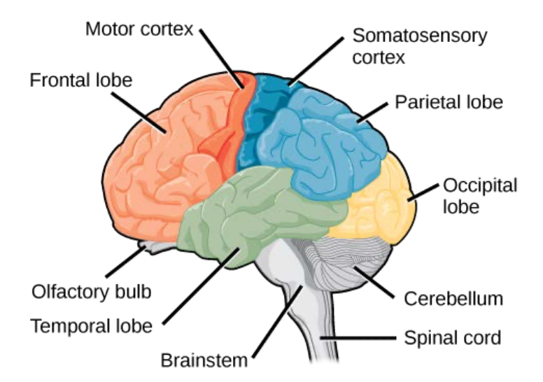
Brain Anatomy Royalty Free Cliparts Vectors And Stock Illustration Image 20850844 from previews.123rf.com Cerebrovascular disease (stroke or brain attack): This amazing organ acts as a control center by receiving, interpreting, and directing sensory information throughout the body. The midbrain helps control eye movement and processes visual and. Humans are born with relatively immature brains that continue to develop in size, shape, and structure throughout childhood and adolescence. The top image shows the four main sections of the cerebral cortex: On the left a coronal view of the segments of the middle cerebral artery. It consists of three structures: It consists of three major parts:
Reviewed by john morrison, patrick hof, and edward lein.
It serves as a relay station, passing messages back and forth between various parts of the body and the cerebral cortex. Map of the human brain: The mapping effort subdivided each half of the brain into 180 separate partitions, with 83 of those already well established in the field, and 97 new areas ripe for exploration. The brainstem is the lower extension of the brain, located in front of the cerebellum and connected to the spinal cord. Large and colourful brain anatomy poster showing the lobes, arteries and cranial nerves. Let's use a common method and divide the brain into three main regions based on embryonic development: The following table shows the brain regions from the cortex to the brain stem with their functions and associated disorders in english, german and latin. The user can manipulate the brain on screen, the app also allows sectioned views and the ability to add custom labels. The brain and spinal cord are the two main structures of the central nervous system. Reviewed by john morrison, patrick hof, and edward lein. The cerebrum, the largest part of the human brain, is associated with higher order functioning, including the control of voluntary behavior. £14.40 inc vat £12.00 exc vat. Structure of the rat brain is presented here as an open access internet resource for the neuroscience community.
Structure of the rat brain is presented here as an open access internet resource for the neuroscience community anatomy map. The mapping effort subdivided each half of the brain into 180 separate partitions, with 83 of those already well established in the field, and 97 new areas ripe for exploration.


Post a Comment